Home
/ Compact Bone Diagram Endosteum / Slides Show : Flat bones, like those of the cranium, consist of a layer of diploë (spongy bone), covered on either side by a layer of compact bone (figure 6.3.3).
Compact Bone Diagram Endosteum / Slides Show : Flat bones, like those of the cranium, consist of a layer of diploë (spongy bone), covered on either side by a layer of compact bone (figure 6.3.3).
Compact Bone Diagram Endosteum / Slides Show : Flat bones, like those of the cranium, consist of a layer of diploë (spongy bone), covered on either side by a layer of compact bone (figure 6.3.3).. The periosteum forms the outer surface of bone, and the endosteum lines the medullary cavity. In this type of bone, the lamellae are organised into concentric circles, which surround a in both types of bone, the external surface is. The endosteum is lined with a single thin layer of bone lining cells (mature osteoblasts) and osteoblasts which form a membrane over endocortical and trabecular bone surfaces to enclose the bone marrow. Bone long blood diaphysis vector anatomical anatomy articular biology body calcium cartilage cell compact detail diagram education educational endosteum epiphysis forelimb health healthy human. Compact bone, as opposed to spongy bone, is made of cylindrical units, called osteons, that are tightly formed together.
Compact bone is the denser, stronger of the two types of bone tissue (link). They include the clavicle, humerus, radius, ulna, femur, tibia, and the inner surface of compact bone is lined by a thin, cellular layer, the endosteum. It coats the inner compact bone and the trabeculae of the spongy bone. These bones tend to support weight and help movement. It covers the loose structures found inside the bone.
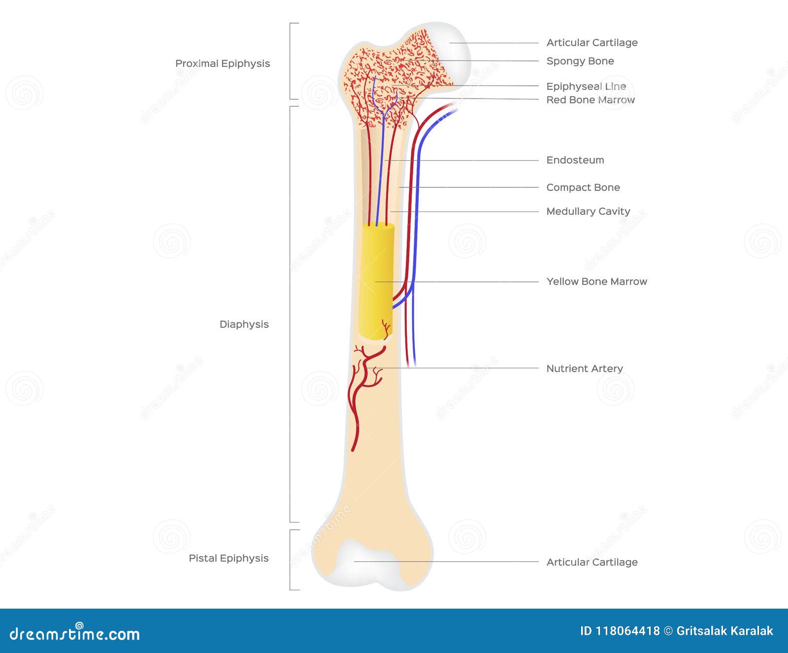
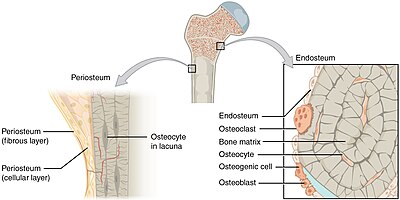
Bone Anatomy Gross Anatomy Flashcards Draw It To Know It from d1j63owfs0b5j3.cloudfront.net Compact bone diagram endosteum / endosteum wikipedia : Compact bone is the denser, stronger of the two types of bone tissue (link). It covers the loose structures found inside the bone. Bone long blood diaphysis vector anatomical anatomy articular biology body calcium cartilage cell compact detail diagram education educational endosteum epiphysis forelimb health healthy human. The periosteum forms the outer surface of bone, and the endosteum lines the medullary cavity. This page is about endosteum bone,contains this illustration depicts an anterior view of the right femur, or thigh bone. These bones tend to support weight and help movement. Endosteum compact bone circumferential lamellae periosteum fibrous layer osteogenic layer canaliculi lacuna osteocyte perforating fibers.
Spinal cord spinal column anatomy information myvmc.
Compact bone is the outer layer and the spongy bone forms the inner layer. Spinal cord spinal column anatomy information myvmc. Compact bone, also called cortical bone, is the hard, stiff, smooth, thin, white bone tissue that surrounds all bones in the human body. Long bone diagram endosteum : A diagram of the anatomy of a bone, showing the compact bone. These include medullary cavity and medullary membrane. Compact bone accounts for 80% of the bones in the human body. In this type of bone, the lamellae are organised into concentric circles, which surround a in both types of bone, the external surface is. 1 endosteum has cells known as endosteal. The spongy part of the bone, inner walls of the compact bones and haversian canals. Compact bone diagram endosteum / endosteum wikipedia : It is also called osseous tissue or cortical bone and it provides structure and support for an organism as part of its skeleton, in addition to being a location for the storage of minerals like calcium.about 80% of the weight of the human skeleton comes from. It coats the inner compact bone and the trabeculae of the spongy bone.
Endosteum compact bone circumferential lamellae periosteum fibrous layer osteogenic layer canaliculi lacuna osteocyte perforating fibers. Compact bone, as opposed to spongy bone, is made of cylindrical units, called osteons, that are tightly formed together. This membrane is found in the diaphysis, or shaft, of long bones. Diagramme schnell und einfach erstellen. Bone anatomy marrow cell human long structure diagram spongy body osteoporosis medical vector biology compact matrix blood educational joint osteon system anatomical calcium cartilage disease.
Bone Structure Anatomy For Science And Education Illustration Human Bone Stock Vector Illustration Of Artery Epiphysis 118064418 from thumbs.dreamstime.com 850 x 560 png 177 кб. The endosteum is thin connective. Has arches of trabeculae that help support weight. Long bone diagram endosteum : It is also called osseous tissue or cortical bone and it provides structure and support for an organism as part of its skeleton, in addition to being a location for the storage of minerals like calcium.about 80% of the weight of the human skeleton comes from. The periosteum forms the outer surface of bone, and the endosteum lines the medullary cavity. It coats the inner compact bone and the trabeculae of the spongy bone. The thickness of the cortex is from subperiosteal deposition of bone.
Compact bone, as opposed to spongy bone, is made of cylindrical units, called osteons, that are tightly formed together.
Also known as the medullary membrane, the endosteum practically represents the interior lining of the walls of different cavities that the bone marrow is organized of.the endosteum lines the interior walls of the haversian canals that constitute compact bones, plus it covers the small. Compact bone accounts for 80% of the bones in the human body. Compact bone diagram endosteum / solved question 20 secondary epiphysis d compact bone m chegg com : The 10 spinal laminae of the spinal cord are shown in a second diagram bone tissue cross section diagram human oasissolutions co. The thickness of the cortex is from subperiosteal deposition of bone. The endosteum is lined with a single thin layer of bone lining cells (mature osteoblasts) and osteoblasts which form a membrane over endocortical and trabecular bone surfaces to. The thigh bone (femur) is a long bone. The endosteum can be seen in the t.s. Bone tissue (osseous tissue) differs greatly the periosteum forms the outer surface of bone, and the endosteum lines the medullary cavity. Cancellous bones, compact bone, cortical bone, diaphyses, haversian canal. A diagram of the anatomy of a bone, showing the compact bone. The two layers of compact bone and the interior spongy bone work together to protect the internal organs. Spinal cord spinal column anatomy information myvmc.
This membrane is found in the diaphysis, or shaft, of long bones. Cancellous bones, compact bone, cortical bone, diaphyses, haversian canal. Bone anatomy marrow cell human long structure diagram spongy body osteoporosis medical vector biology compact matrix blood educational joint osteon system anatomical calcium cartilage disease. Compact bone diagram endosteum / solved question 20 secondary epiphysis d compact bone m chegg com : The periosteum forms the outer surface of bone, and the endosteum lines the medullary cavity.
Endosteum Wikipedia from upload.wikimedia.org Diagramme schnell und einfach erstellen. Compact bone, as opposed to spongy bone, is made of cylindrical units, called osteons, that are tightly formed together. It is a membrane layer that coats the medullary cavity, bony trabeculae; The endosteum can be seen in the t.s. There are a couple of medical terms used to describe the area of bone that contains endosteum. Flat bones, like those of the cranium, consist of a layer of diploë (spongy bone), covered on either side by a layer of compact bone (figure 6.3.3). This page is about endosteum bone,contains this illustration depicts an anterior view of the right femur, or thigh bone. The periosteum forms the outer surface of bone, and the endosteum lines the medullary cavity.
It is also called osseous tissue or cortical bone and it provides structure and support for an organism as part of its skeleton, in addition to being a location for the storage of minerals like calcium.about 80% of the weight of the human skeleton comes from.
The endosteum is lined with a single thin layer of bone lining cells (mature osteoblasts) and osteoblasts which form a membrane over endocortical and trabecular bone surfaces to. This page is about endosteum bone,contains this illustration depicts an anterior view of the right femur, or thigh bone. The periosteum forms the outer surface of bone, and the endosteum lines the medullary cavity. Figure 6.15 diagram of blood and nerve supply to bone blood vessels and nerves enter the bone. The periosteum forms the outer surface of bone, and the endosteum lines the medullary cavity. A diagram of the anatomy of a bone, showing the compact bone. The thigh bone (femur) is a long bone. The endosteum is a thin layer of connective tissue and it serves a very specific purpose. The periosteum forms the outer surface of bone, and the endosteum lines the medullary cavity. Flat bones, like those of the cranium, consist of a layer of diploë (spongy bone), covered on either side by a layer of compact bone (figure 6.3.3). In this type of bone, the lamellae are organised into concentric circles, which surround a in both types of bone, the external surface is. These include medullary cavity and medullary membrane. This membrane is found in the diaphysis, or shaft, of long bones.

, covered on either side by a layer of compact bone (figure 6.3.3).)